


This article is a submission from the NeuroNYC community, edited by the NeuroNYC team. The content does not represent the official views of NeuroNYC, nor is it meant as an endorsement of mentioned groups. We encourage community submissions: please email your name, affiliation (if any), and short pitch for your article to hello@neuro-nyc.org.
The idea behind a brain–computer interface is straightforward in principle: record neural activity, decode it into commands, and use those commands to drive a digital device: a cursor, a robotic arm, a speech synthesizer, etc. The field is now moving beyond pure readout. Newer systems use microstimulation to write information back into the brain, and some close the loop entirely with bidirectional implants that sense and stimulate within the same device.
But before any of this reaches a person, every layer of the system has to be validated: whether the electrodes couple to tissue, whether the signals are genuinely neural, and whether they carry enough information to be useful. This post walks through that validation stack using recent preclinical publications from Precision Neuroscience1, which makes a thin-film surface array, and Paradromics2, which makes a high-density intracortical array.
1. Bench signal tests and impedance
Before an electrode records neurons, engineers need to know how well it couples to tissue. The measure for this is electrode impedance. Impedance demonstrates how strongly the electrode–tissue interface resists alternating current, and it is usually reported at 1 kHz, roughly the frequency band of an action potential.
Impedance matters for three reasons:
Noise. Thermal noise rises with impedance. The noise of a high-impedance site can bury small neural signals, which are often only tens of microvolts, under its own noise floor.
Size vs. resolution. Smaller electrodes see more local activity but have higher impedance. Every array design has to balance spatial resolution against signal quality.
Health monitoring. Impedance that drifts over time can warn of trouble: a broken trace, failing insulation, or scar tissue forming around the implant. Teams measure it before implantation, right after, and throughout chronic studies.
Precision’s minipig study shows the size trade-off clearly. Average 1 kHz impedance ran from about 802 kΩ for 20 µm electrodes to 8.25 kΩ for 380 µm electrodes, and impedance changed little from before to after implantation1. That second result shows the ultra-thin film survived being slid onto the cortical surface.
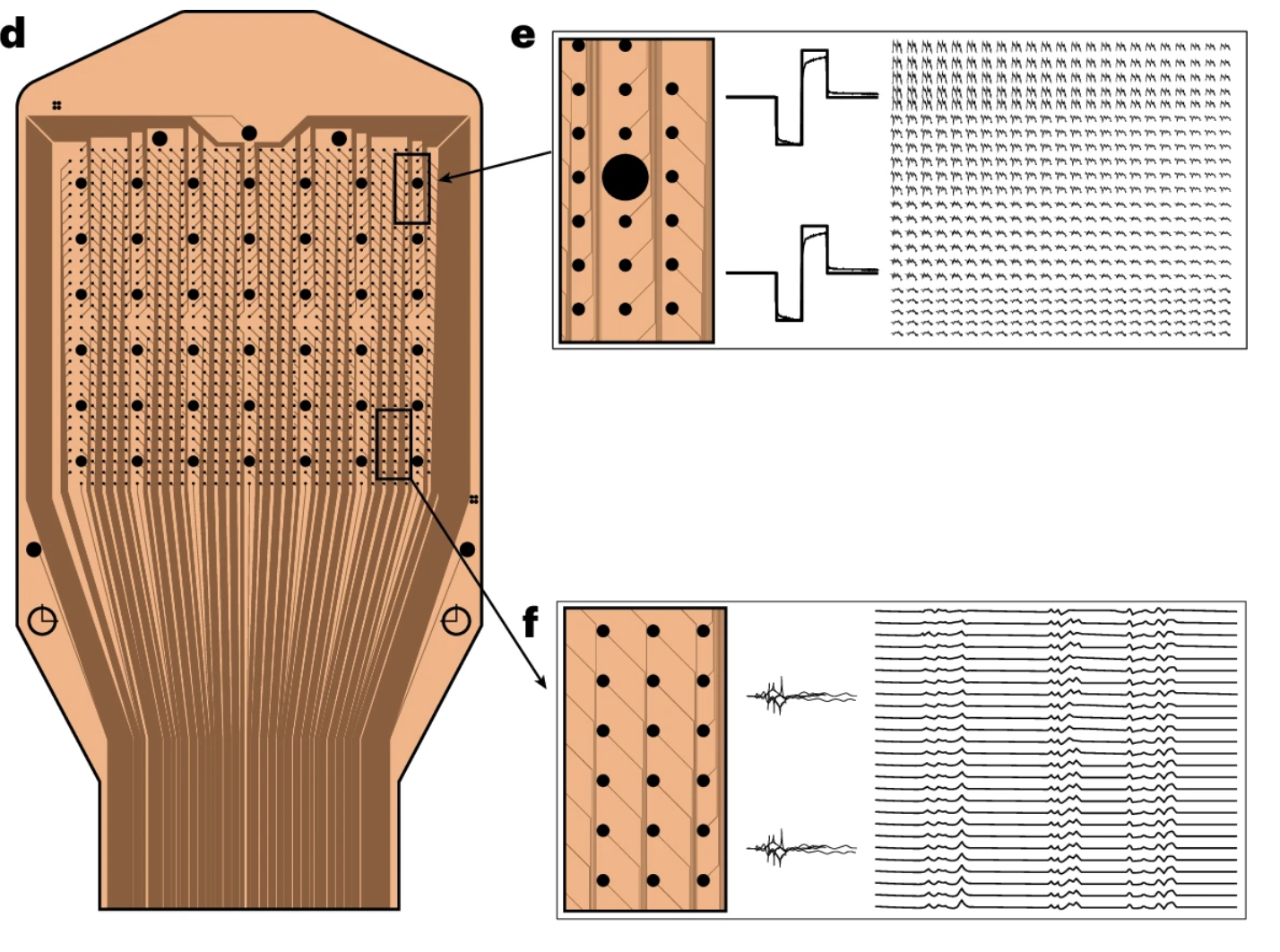
Top: 380 µm electrodes, bottom: 50 µm electrodes
2. Neural signal characterization
A low-impedance electrode only proves the device can pick up something. Next you have to show that the signal is coming from neurons and not from noise, line interference, or movement artifacts. Neural activity has a few physical signatures that can be tested for.
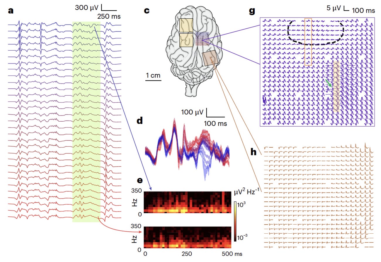
Spatial structure. Dense electrode arrays record the local field potential (LFP), the summed electrical activity of nearby neural populations. Because neighboring electrodes sample overlapping populations, their signals should be correlated, and that correlation should fall off with distance. Precision reported beta band r² of 0.67 between 50 µm electrodes spaced 300 µm apart, with correlation dropping faster at higher frequencies1. That pattern fits a real cortical source. However, a movement artifact would look roughly the same on every channel.
Stimulus-locked responses. When the brain receives a known input, the recording should capture its timely response. Somatosensory-evoked potentials (SEPs) and visual-evoked potentials (VEPs) have well-known timing and anatomy. They should appear at a consistent delay after the stimulus and over the expected patch of cortex. In Precision’s Göttingen minipigs, touching the hindlimb produced SEPs over somatosensory cortex that were organized in both time and space across the array1. Flashing light into each eye separately produced VEPs over visual cortex.
3. BCI Decoding
A clean, neural signal is still only a starting point. A BCI is a communication channel, so its performance comes down to how much true information gets from the brain to the computer, and how quickly. Three things set that ceiling:
Recording quality: channel count, signal-to-noise ratio (how cleanly the channels picks up neural signals relative to background noise), and channel placement (ideally over a cortical area that responds to the task).
The decoder: the algorithm that translates raw neural signals into a predicted output. This can be anything from linear models and template matching to deep networks.
The task: what the brain is being asked to communicate, and how much information that involves.
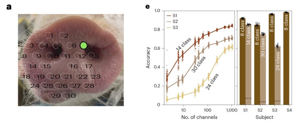
Precision tested multi-point somatosensory discrimination using a tactile stimulation paradigm in minipigs. Their 1024-channel array was placed over somatosensory cortex, and the animal’s rostrum was stimulated at predefined locations in a randomized sequence. Using a 1D convolutional neural network (CNN) combined with bidirectional gated recurrent units (biGRUs), the team decoded stimulation site from neural activity with a mean accuracy of 92.75%1.
Paradromics built a dedicated benchmark called SONIC (Standard for Optimizing Neural Interface Capacity). The Connexus device is implanted in sheep auditory cortex, which is organized tonotopically so that different cortical regions respond preferentially to different sound frequencies. The team plays 10ms tones in a random sequence to live sheep and measures how much information about the tone played is preserved in what the algorithm decodes from the neural activity. A feature extractor paired with a 1D CNN accurately recovered tone frequency from the neural data.
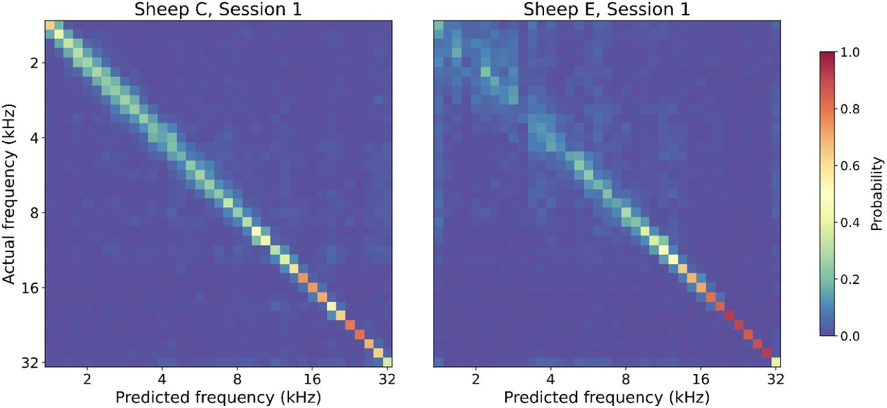
Together, these studies demonstrate BCI implants can decode precisely timed neural information across different sensory modalities.
Towards bedside applications
Scaling a BCI product means more than technical success. Labs and companies are testing the chronic stability and biocompatibility of their devices over months and years. BrainGate has reported Utah Array implants remaining functional in human participants for up to 7.6 years3. On the preclinical side, Paradromics has demonstrated four years of stable, healthy neural recordings from sheep chronically implanted with the Connexus device4.
Access remains another constraint. Getting one of these devices still requires a neurosurgical procedure, which limits who can benefit and where trials can run. The companies that ultimately reach the bedside will be the ones that can show results at every layer of this validation stack, not only in the demo video.
References:
Hettick, M., Ho, E., Poole, A.J. et al. Minimally invasive implantation of scalable high-density cortical microelectrode arrays for multimodal neural decoding and stimulation. Nature Biomedical Engineering 10, 1206–1221 (2026). https://doi.org/10.1038/s41551-025-01501-w
Perkins SM, Trumpis M, Reitman ME, Jarosiewicz B, Patel AN, Weiss A, et al. SONIC: A Benchmarking Paradigm for Brain-Computer Interfaces. Preprint at https://doi.org/10.1101/2025.09.30.679683 (2025).
Hahn, N.V., Stein, E., BrainGate Consortium, Donoghue, J.P., Simeral, J.D., Hochberg, L.R., & Willett, F.R. Long-term performance of intracortical microelectrode arrays in 14 BrainGate clinical trial participants. medRxiv, 2025. https://doi.org/10.1101/2025.07.02.25330310
Paradromics. Four years of stable neural recording milestone. Press release, 2026.
Acknowledgements:
The information and figures from “Minimally invasive implantation of scalable high-density cortical microelectrode arrays for multimodal neural decoding and stimulation” (Hettick et al. 2026), including the article thumbnail, are shared under a CC-BY-NC-ND 4.0 International License: http://creativecommons.org/licenses/by-nc-nd/4.0/. All figures shared have been cropped for specificity.
The information and figure from “SONIC: A Benchmarking Paradigm for Brain–Computer Interfaces” (Perkins et al. 2025) are shared under a CC-BY-NC-ND 4.0 International License: http://creativecommons.org/licenses/by-nc-nd/4.0/.